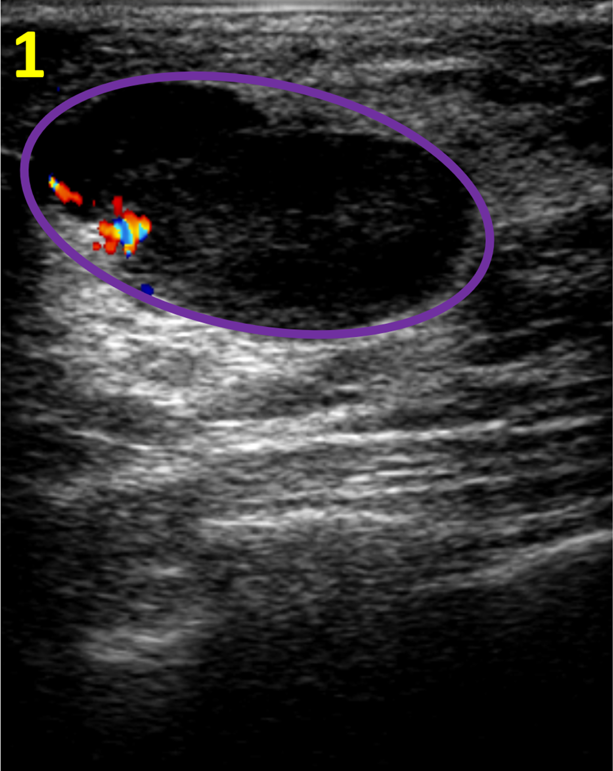
The thought of breast cancer is a frightening one, and it’s natural to feel anxious about any potential signs. One of the tests often used for breast cancer screening is a breast ultrasound. For many, the experience itself can be unnerving, and then there’s the added confusion of seeing the image on the screen, with its mysterious red and blue colors. What do these colors mean and what are they showing you? Today, we’ll delve into the world of breast ultrasounds and unravel the mystery of the red and blue color scheme.

Image: hairstyle.udlvirtual.edu.pe
I’ll never forget the day I received my ultrasound results. The technician handed me the image, and I stared at it, puzzled. It was a mosaic of red, blue, and shades of grey, and I had no idea what it all meant. I quickly started Googling, but the information I found was too technical, and I just felt more confused. I wish I had had a guide like this one that explained the colors and their importance in a clear and understandable way.
Understanding the Red and Blue Spectrum
When you look at a breast ultrasound image, you’re actually seeing a cross-sectional view of your breast tissue. The different colors represent different densities of tissue. Red and blue are often used in these images to delineate different types of tissue and indicate areas of concern, though exact colors can vary based on the specific machine used.
Ultrasound images are created by sending high-frequency sound waves into the body and then analyzing the echoes that bounce back. Denser tissues, like tumors, tend to reflect more sound waves, creating a brighter and often redder color on the image, while less dense tissues, like fat, absorb more sound and appear darker, often as blues or shades of grey.
Delving Deeper into the Meaning of Colors and Their Significance
Red: Potential Concerns
Red areas, often the brightest on the image, can indicate dense, abnormal tissue that requires closer examination. This does not mean it’s cancer, as cysts and other benign conditions can also appear red. However, it is a red flag for the physician and requires further examination.

Image: www.cureus.com
Blue and Grey: Relatively Normal Tissues
Blue and shades of grey typically represent less dense tissues, including normal breast tissue, fat, and other fluids. They may not be as visually striking as red areas, but they play a crucial role in understanding the complex structure of the breast.
How the Colors Aid in Diagnosis
The red and blue color scheme aids in diagnosing breast issues by highlighting areas of concern but understanding the color is only one aspect of the diagnosis.
A radiologist, trained in interpreting ultrasound images, evaluates the entire image, seeking patterns, shapes, and textures that might indicate a problem. They also consider your medical history, age, and any other relevant factors before making a diagnosis.
Key Tips and Expert Advice for Understanding your Ultrasound Results
Remember that an Ultrasound is Just One Piece of the Puzzle
An ultrasound is a valuable tool, but it’s just one part of a comprehensive evaluation. There are other imaging tests, biopsies, and physical exams to consider. Always consult with your doctor before drawing any conclusions based solely on your ultrasound image.
Do Not Interpret Your Ultrasound on Your Own
It’s tempting to try to interpret your ultrasound image yourself, especially with the plethora of information available online, but resist this urge! Only a qualified radiologist can accurately interpret these images and provide you with the right information and guidance.
Don’t Forget the Importance of Regular Screenings
The best way to catch potential breast cancer early is to have regular screenings, including mammograms, clinical breast exams, and ultrasounds, as recommended by your doctor. Early detection dramatically improves the odds of successful treatment.
FAQ about Breast Cancer and Ultrasound Examinations
Q: Is an ultrasound for breast cancer painful?
A: Most people find ultrasounds to be painless. You may feel a slight pressure from the transducer, but there should be no pain.
Q: How often should I get a breast ultrasound?
A: The frequency of recommended breast ultrasounds varies based on individual risk factors, family history, and age. Your doctor can provide personalized guidance on your screening needs.
Q: What if I see red areas on my ultrasound image?
A: If you notice red areas on your ultrasound image, don’t panic! This doesn’t automatically mean you have cancer. Your radiologist will assess the entire image and determine the cause of the red color. They will guide you through the next steps based on their interpretation.
Breast Cancer Breast Ultrasound Red And Blue Color
https://youtube.com/watch?v=ODVWAijqcS4
Conclusion
Understanding the red and blue colors on a breast ultrasound can help you feel more informed and empowered. Remember, these colors don’t provide a definitive diagnosis; they’re just a tool to highlight potential areas of concern. Your doctor is the best resource for interpreting your results and finding the best course of action for your specific situation. It’s a good habit to follow up with any questions or concerns you may have, empowering you to take control of your health journey.
Are you curious about breast cancer ultrasounds or have any questions about the technology?


