Have you ever peered through a microscope and wondered why the nucleus of a cell appears a vibrant shade of purple or blue? The answer lies within the fascinating world of microscopy and the intricate dance between stains and cellular components. Understanding why the nucleus stains a specific color during microscopic observation allows scientists to delve deeper into the inner workings of cells, revealing their secrets and advancing our knowledge of biology.

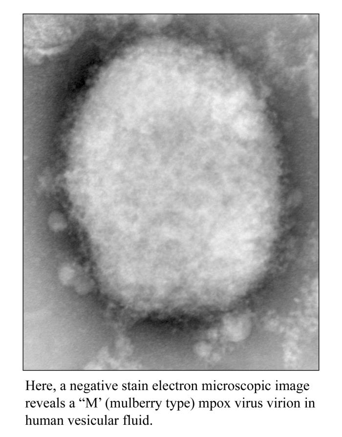
Image: phil.cdc.gov
The nucleus, often referred to as the “brain” of the cell, is the control center that houses the cell’s DNA. This genetic material directs all cellular activities, from protein synthesis to cell division. The nucleus is a dynamic structure, constantly working to ensure the cell functions properly, making it a key target for scientific investigation. Microscopy, the art of magnifying tiny objects beyond the limits of the naked eye, provides us with the tools to investigate the intricate details of cells, including their nucleus.
The Magic of Stains
To visualize the nucleus under a microscope, scientists employ a technique called staining. Staining involves using dyes that bind selectively to specific cellular structures, enhancing their visibility and enabling researchers to differentiate between various components. This process is akin to painting a complex canvas where each color represents a unique cellular structure. The nucleus, with its high concentration of DNA, is particularly adept at absorbing certain stains, revealing its intricate structure under the microscope.
A Love Affair with Basic Dyes
The most common stains used to highlight the nucleus are basic dyes. These dyes possess a positive charge, making them attracted to the negatively charged phosphate groups found in DNA. This attraction is what makes the nucleus “light up” against the rest of the cell, making it readily visible.
One of the most popular basic dyes is methylene blue, which imparts a beautiful blue hue to the nucleus. Another commonly used stain is hematoxylin, a natural dye extracted from the heartwood of the logwood tree. Hematoxylin, when used in conjunction with a mordant (a substance that helps the dye bind), stains the nucleus a vivid purple.
The Dance of Hematoxylin and Eosin
In histology, a branch of biology that focuses on the study of tissues, the combo of hematoxylin and eosin (a red dye) reigns supreme. This duo creates stunningly contrasting colors, highlighting the nucleus in purple and the cytoplasm (the jelly-like substance that surrounds the nucleus) in a delicate pink. This combination, known as H&E staining, is widely used in diagnostic pathology to identify various diseases and abnormalities in tissues.

Image: www.snaggledworks.com
Why the Nucleus Stains: A Deeper Dive
The nucleus’s affinity for basic dyes boils down to its chemical composition. DNA, the blueprint of life, is a long, spiraling molecule composed of repeating units called nucleotides. Each nucleotide consists of a sugar molecule, a phosphate group, and a nitrogenous base. The phosphate groups, with their negative charges, attract the positively charged basic dyes, leading to the nucleus’s vibrant staining.
Variations in Nuclear Staining
While the nucleus generally stains with basic dyes, variations in staining intensity can occur between cells depending on factors such as:
-
Cell type: Different cell types may have varying amounts of DNA, leading to differences in staining intensity. For example, cells actively engaged in protein synthesis often have a more prominent and intensely stained nucleus.
-
Cell cycle: The stage of the cell cycle can also influence nuclear staining. During cell division, the nucleus replicates its DNA, resulting in a more intensely stained nucleus.
-
Cellular activity: Cells that are metabolically active, such as nerve cells and muscle cells, often exhibit a larger and more intensely stained nucleus due to their higher DNA content.
Unlocking the Secrets of the Nucleus
The fact that the nucleus stains a specific color during microscopic observation is a testament to the powerful tools scientists have at their disposal. By understanding the chemical properties of both the nucleus and the stains, researchers can identify specific features of cells and gain insights into their function. This knowledge has numerous applications, including:
-
Disease diagnosis: Pathologists use microscopic observation and staining techniques to identify cancerous cells and other abnormalities in tissues. The nucleus, with its telltale purple hue, serves as a critical marker for diagnosis.
-
Drug development: Pharmaceutical companies utilize microscopy and staining to test the effects of potential drugs on cells. By studying nuclear changes, they can assess the efficacy and safety of new pharmaceuticals.
-
Environmental monitoring: Scientists use microscopic analysis to assess the impact of environmental toxins on cells. The nucleus, often a primary target for toxic substances, can indicate the severity of environmental damage.
Beyond the Microscope: The Future of Nuclear Staining
While traditional staining techniques are still widely used, the field of microscopy is constantly evolving. Scientists are developing new methods to provide even more detailed insights into the nucleus and its role in cellular function.
-
Fluorescent staining: This technique involves using dyes that emit light upon excitation with specific wavelengths of light. This enables researchers to visualize specific structures within the nucleus, such as chromosomes and nucleoli.
-
Immunofluorescence: Antibodies, proteins that bind to specific targets, are tagged with fluorescent dyes. This technique allows for the visualization of protein structures within the nucleus, providing information about protein function and cellular signaling pathways.
-
Electron microscopy: This advanced technique uses a beam of electrons to create high-resolution images of cells and their internal structures. Electron microscopy provides a much more detailed view of the nucleus and its internal compartments.
Which Color Will The Nucleus Stain During Microscopic Observation
Conclusion
The fact that the nucleus stains a specific color during microscopic observation is much more than just a visual phenomenon. It’s a window into the intricate world of cells, revealing their complex organization and dynamic processes. Through the use of stains, scientists have unlocked a wealth of knowledge about the nucleus, its role in cellular function, and its involvement in various biological processes. As microscopy continues to evolve, we can expect even more groundbreaking discoveries to be made, shedding light on the secrets of the nucleus and the fascinating world of cells.


